Terminology and Classification
Choanal atresia is secondary to the unilateral or bilateral persistence of the buccopharyngeal membrane. It can be membranous or bony. It is important to define how thick any bony stenosis is and study the anatomy of neighboring structures such as the carotid artery.
Indications
A newborn infant relies on its nasal airway. It is particularly compromised during suckling. It is not possible to leave bilateral choanal atresia untreated. Rapid assessment and correction are needed to provide a nasal airway, and an oropharyngeal airway has to be held in place in the meantime. Bilateral choanal atresia is often associated with other congenital abnormalities (the CHARGE syndrome in particular) that also need to be investigated.
First of all an attempt to pass a fine nasogastric tube into the nasopharynx should be made to help confirm the diagnosis. An axial CT should be done, but only after decongesting the mucosa and sucking any mucus (this avoids mucus producing the false appearance of a mucosal obstruction). A unilateral atresia often presents later in the early teens when the patient realizes that they cannot breathe through one side. They can also present with a unilateral mucoid discharge.
Surgical Anatomy
A persistent membrane or bony plate separates the nasopharynx from the nasal airway. Not only does the thickness of any bony obstruction need to be assessed but also its lateral and medial intrusion into the airway. There may be no complete bony partition, but thick lateral bone that protrudes medially and narrows the airway will need a lot of bony work in order to widen it.
Surgical Technique
If the obstruction is due to soft tissue then it is possible to palpate it and see with an endoscope where it should be perforated and opened up. If there is a complete bony plate, it is important not to lose your way, and most surgeons initially find it safer to place their finger in the nasopharynx and aim the drill at it. It is now possible to visualize the posterior aspect of the septum and the back of the inferior turbinate and use these landmarks to drill through the atretic plate under endoscopic control. Having made an opening by whatever means, it can then be widened endoscopically in a controlled way. The primary goals of surgery are to provide a wide airway with as little collateral mucosal damage as possible.
Conventionally, a wide stent has been inserted, but this causes pressure necrosis of any viable mucosa and it appears to encourage fibrosis and stenosis once the stent is removed. A loose stent is better, and the strut joining the two cylindrical stents should not press on the columella. An endotracheal tube that has a section cut out of it to leave one flat connecting piece between the two tubes can lightly rest on the columella with a loose circumferential tie placed through the tubes and around the nasopharynx to retain it.
Unilateral Atresia
In unilateral choanal atresia a simple technique that allows aeration and restoration of mucociliary clearance of the blocked airway relies on removing the vomer. This can be done endoscopically by incising through all layers just behind the quadrilateral cartilage and then removing all layers of the vomer with through-cutting forceps (Cumberworth et al., 1995). The septal branch of the sphenopalatine artery usually needs to be cauterized. No stent is required.
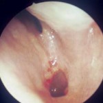
- Preoperative view of choanal atresia
- Postoperative view
a Axial CT scan showing bilateral choanal atresia. b The right nasal cavity showing that the back of the vomer has been removed as well as the floor of the sphenoid sinus. c Postoperative view into the sphenoid and into the oropharynx below.
- a bilateral choanal atresia
- b right nasal cavity during operation
- c after operation
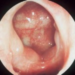
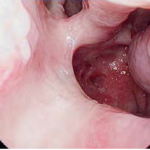
a Mucus stagnation in unilateral choanal atresia. b Axial CT scan showing a left unilateral membranous choanal atresia. c Postoperative endoscopic view showing a patent posterior choana 8 years later.
- a Mucus stagnation in unilateral choanal atresia
- b left unilateral membranous choanal atresia
- c Postoperative endoscopic view
Alternative Surgical Techniques
Transpalatal techniques allow direct exposure but cause more disruption and fibrosis of the palatal muscles.


